Main Article Content
Abstract
Background:
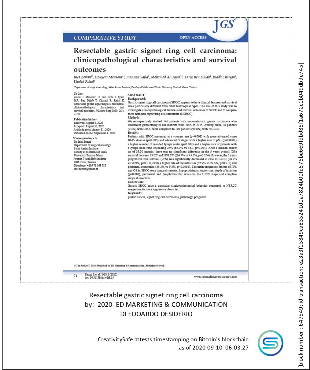
Gastric signet ring cell carcinoma (SRCC) appears to have clinical features and survival rates particularly different from other histological types. The aim of this study was to investigate clinicopathological features and survival outcomes of SRCC and to compare them with non-signet ring cell carcinoma (NSRCC).
Methods:
We retrospectively studied 145 patients with non-metastatic gastric carcinoma who underwent gastrectomy in our institute from 2005 to 2015. Among them, 36 patients (9.4%) with SRCC were compared to 109 patients (90.6%) with NSRCC.
Results:
Patients with SRCC presented at a younger age (p=0.001) with more advanced stage III-IV disease (p=0.005) and advanced N stages with a higher rate of pN3 (p=0.0001), a higher number of invaded lymph nodes (p=0.002) and a higher rate of patients with a lymph node ratio exceeding 25% (63.9% vs 36.7, p=0.004). After a median follow up of 35.30 months, there was no significant difference in the 5 years overall (OS) survival between SRCC and NSRCC ((36.7% vs 45.7%, p=0.206).However, the 5 years progressive free survival (PFS) was significantly decreased in case of SRCC (38.7% vs 50.9%, p=0.038) with a higher rate of metastasis in (52.9% vs 29.5%, p=0.013) and peritoneal recurrence (35.3% vs 9.5%, p<0.0001). The main prognostic factors of PFS and OS in SRCC were tumoral stenosis, hypoprotidemia, tumor size, depth of invasion (p=0.001), perineural and lymphovascular invasion, the UICC stage and complete surgical resection.
Conclusion:
Gastric SRCC have a particular clinicopathological behavior compared to NSRCC suggesting its more aggressive character.
Keywords
Article Details
References
- Hass HG, Smith U, Jäger C, et al. Signet ring cell carcinoma of the stomach is significantly associated with poor prognosis and diffuse gastric cancer (Lauren’s): single-center experience of 160 cases. Oncology Research and Treatment. 2011; 34: 682-6.
- Chu PG and Weiss LM. Immunohistochemical characterization of signet-ring cell carcinomas of the stomach, breast, and colon. American journal of clinical pathology. 2004; 121: 884-92.
- Lauren P. The two histological main types of gastric carcinoma: diffuse and so‐called intestinal‐type carcinoma: an attempt at a histo‐clinical classification. Acta Pathologica Microbiologica Scandinavica. 1965; 64: 31-49.
- Nakamura T and Nakano G. Histopathological classification and malignant change in gastric polyps. Journal of clinical pathology. 1985; 38: 754-64.
- Lee HH, Song KY, Park CH and Jeon HM. Undifferentiated-type gastric adenocarcinoma: prognostic impact of three histological types. World journal of surgical oncology. 2012; 10: 254.
- Ćerban D, Brănescu C, Ćavlovschi C, et al. Particularităĺi clinico-morfologice într-un caz de carcinom gastric incipient (tipul de carcinom în inel cu pecete). Chirurgia. 2011; 106: 383-7.
- Taghavi S, Jayarajan SN, Davey A and Willis AI. Prognostic significance of signet ring gastric cancer. Journal of clinical oncology. 2012; 30: 3493.
- Matsuyama S, Ohkura Y, Eguchi H, et al. Estrogen receptor β is expressed in human stomach adenocarcinoma. Journal of cancer research and clinical oncology. 2002; 128: 319-24.
- Li C, Kim S, Lai JF, et al. Advanced gastric carcinoma with signet ring cell histology. Oncology. 2007; 72: 64-8.
- Nie R-C, Yuan S-Q, Li Y-F, et al. Clinicopathological characteristics and prognostic value of signet ring cells in gastric carcinoma: A meta-analysis. Journal of Cancer. 2017; 8: 3396.
- Piessen G, Messager M, Leteurtre E, Jean-Pierre T and Mariette C. Signet ring cell histology is an independent predictor of poor prognosis in gastric adenocarcinoma regardless of tumoral clinical presentation. Annals of surgery. 2009; 250: 878-87.
- Kwon K-J, Shim K-N, Song E-M, et al. Clinicopathological characteristics and prognosis of signet ring cell carcinoma of the stomach. Gastric Cancer. 2014; 17: 43-53.
- Chen Y-C, Fang W-L, Wang R-F, et al. Clinicopathological variation of Lauren classification in gastric cancer. Pathology & Oncology Research. 2016; 22: 197-202.
- Kunisaki C, Shimada H, Nomura M, Matsuda G, Otsuka Y and Akiyama H. Therapeutic strategy for signet ring cell carcinoma of the stomach. British journal of surgery. 2004; 91: 1319-24.
- Jiang CG, Wang ZN, Sun Z, Liu FN, Yu M and Xu HM. Clinicopathologic characteristics and prognosis of signet ring cell carcinoma of the stomach: Results from a chinese mono‐institutional study. Journal of surgical oncology. 2011; 103: 700-3.
- Gronnier C, Messager M, Robb WB, et al. Is the negative prognostic impact of signet ring cell histology maintained in early gastric adenocarcinoma? Surgery. 2013; 154: 1093-9.
- Kim DY, Park YK, Joo JK, et al. Clinicopathological characteristics of signet ring cell carcinoma of the stomach. ANZ journal of surgery. 2004; 74: 1060-4.
- Liu X, Cai H, Sheng W, et al. Clinicopathological characteristics and survival outcomes of primary signet ring cell carcinoma in the stomach: retrospective analysis of single center database. PloS one. 2015; 10: e0144420.
- Voron T, Messager M, Duhamel A, et al. Is signet-ring cell carcinoma a specific entity among gastric cancers? Gastric Cancer. 2016; 19: 1027-40.
- Zhang M, Zhu G, Zhang H, Gao H and Xue Y. Clinicopathologic features of gastric carcinoma with signet ring cell histology. Journal of Gastrointestinal Surgery. 2010; 14: 601-6.
- Postlewait LM, Squires MH, Kooby DA, et al. The prognostic value of signet-ring cell histology in resected gastric adenocarcinoma. Annals of surgical oncology. 2015; 22: 832-9.
- Song KY, Park YG, Jeon HM and Park CH. A nomogram for predicting individual survival of patients with gastric cancer who underwent radical surgery with extended lymph node dissection. Gastric Cancer. 2014; 17: 287-93.
- Han D-S, Suh Y-S, Kong S-H, et al. Nomogram predicting long-term survival after d2 gastrectomy for gastric cancer. Journal of clinical oncology : official journal of the American Society of Clinical Oncology. 2012; 30: 3834-40.
- Kim DH, Kim SM, Hyun JK, et al. Changes in postoperative recurrence and prognostic risk factors for patients with gastric cancer who underwent curative gastric resection during different time periods. Annals of surgical oncology. 2013; 20: 2317-27.
- Kim J-P, Kim Y-W, Yang H-K and Noh D-Y. Significant prognostic factors by multivariate analysis of 3926 gastric cancer patients. World journal of surgery. 1994; 18: 872-7.
- Wang X, Wan F, Pan J, Yu GZ, Chen Y and Wang JJ. Tumor size: A non‐neglectable independent prognostic factor for gastric cancer. Journal of surgical oncology. 2008; 97: 236-40.
- Saito H, Osaki T, Murakami D, et al. Macroscopic tumor size as a simple prognostic indicator in patients with gastric cancer. The American journal of surgery. 2006; 192: 296-300.
- Xue L, Chen X-L, Lin P-P, et al. Impact of capillary invasion on the prognosis of gastric adenocarcinoma patients: A retrospective cohort study. Oncotarget. 2016; 7: 31215.
- Del Casar JM, Corte MD, Álvarez A, et al. Lymphatic and/or blood vessel invasion in gastric cancer: relationship with clinicopathological parameters, biological factors and prognostic significance. Journal of cancer research and clinical oncology. 2008; 134: 153.
- Selcukbiricik F, Tural D, Buyukunal E and Serdengecti S. Perineural invasion independent prognostic factors in patients with gastric cancer undergoing curative resection. Asian Pacific journal of cancer prevention : APJCP. 2012; 13: 3149-52.
- Liebig C, Ayala G, Wilks JA, Berger DH and Albo D. Perineural invasion in cancer: a review of the literature. Cancer: Interdisciplinary International Journal of the American Cancer Society. 2009; 115: 3379-91.
- Deng J, You Q, Gao Y, et al. Prognostic value of perineural invasion in gastric cancer: a systematic review and meta-analysis. PloS one. 2014; 9: e88907.
- Pernot S, Voron T, Perkins G, Lagorce-Pages C, Berger A and Taieb J. Signet-ring cell carcinoma of the stomach: Impact on prognosis and specific therapeutic challenge. World Journal of Gastroenterology: WJG. 2015; 21: 11428.
- Zheng H-L, Lu J, Li P, et al. Effects of preoperative malnutrition on short-and long-term outcomes of patients with gastric cancer: can we do better? Annals of surgical oncology. 2017; 24: 3376-85.
- Crumley AB, Stuart RC, McKernan M and McMillan DC. Is hypoalbuminemia an independent prognostic factor in patients with gastric cancer? World journal of surgery. 2010; 34: 2393-8.
References
Hass HG, Smith U, Jäger C, et al. Signet ring cell carcinoma of the stomach is significantly associated with poor prognosis and diffuse gastric cancer (Lauren’s): single-center experience of 160 cases. Oncology Research and Treatment. 2011; 34: 682-6.
Chu PG and Weiss LM. Immunohistochemical characterization of signet-ring cell carcinomas of the stomach, breast, and colon. American journal of clinical pathology. 2004; 121: 884-92.
Lauren P. The two histological main types of gastric carcinoma: diffuse and so‐called intestinal‐type carcinoma: an attempt at a histo‐clinical classification. Acta Pathologica Microbiologica Scandinavica. 1965; 64: 31-49.
Nakamura T and Nakano G. Histopathological classification and malignant change in gastric polyps. Journal of clinical pathology. 1985; 38: 754-64.
Lee HH, Song KY, Park CH and Jeon HM. Undifferentiated-type gastric adenocarcinoma: prognostic impact of three histological types. World journal of surgical oncology. 2012; 10: 254.
Ćerban D, Brănescu C, Ćavlovschi C, et al. Particularităĺi clinico-morfologice într-un caz de carcinom gastric incipient (tipul de carcinom în inel cu pecete). Chirurgia. 2011; 106: 383-7.
Taghavi S, Jayarajan SN, Davey A and Willis AI. Prognostic significance of signet ring gastric cancer. Journal of clinical oncology. 2012; 30: 3493.
Matsuyama S, Ohkura Y, Eguchi H, et al. Estrogen receptor β is expressed in human stomach adenocarcinoma. Journal of cancer research and clinical oncology. 2002; 128: 319-24.
Li C, Kim S, Lai JF, et al. Advanced gastric carcinoma with signet ring cell histology. Oncology. 2007; 72: 64-8.
Nie R-C, Yuan S-Q, Li Y-F, et al. Clinicopathological characteristics and prognostic value of signet ring cells in gastric carcinoma: A meta-analysis. Journal of Cancer. 2017; 8: 3396.
Piessen G, Messager M, Leteurtre E, Jean-Pierre T and Mariette C. Signet ring cell histology is an independent predictor of poor prognosis in gastric adenocarcinoma regardless of tumoral clinical presentation. Annals of surgery. 2009; 250: 878-87.
Kwon K-J, Shim K-N, Song E-M, et al. Clinicopathological characteristics and prognosis of signet ring cell carcinoma of the stomach. Gastric Cancer. 2014; 17: 43-53.
Chen Y-C, Fang W-L, Wang R-F, et al. Clinicopathological variation of Lauren classification in gastric cancer. Pathology & Oncology Research. 2016; 22: 197-202.
Kunisaki C, Shimada H, Nomura M, Matsuda G, Otsuka Y and Akiyama H. Therapeutic strategy for signet ring cell carcinoma of the stomach. British journal of surgery. 2004; 91: 1319-24.
Jiang CG, Wang ZN, Sun Z, Liu FN, Yu M and Xu HM. Clinicopathologic characteristics and prognosis of signet ring cell carcinoma of the stomach: Results from a chinese mono‐institutional study. Journal of surgical oncology. 2011; 103: 700-3.
Gronnier C, Messager M, Robb WB, et al. Is the negative prognostic impact of signet ring cell histology maintained in early gastric adenocarcinoma? Surgery. 2013; 154: 1093-9.
Kim DY, Park YK, Joo JK, et al. Clinicopathological characteristics of signet ring cell carcinoma of the stomach. ANZ journal of surgery. 2004; 74: 1060-4.
Liu X, Cai H, Sheng W, et al. Clinicopathological characteristics and survival outcomes of primary signet ring cell carcinoma in the stomach: retrospective analysis of single center database. PloS one. 2015; 10: e0144420.
Voron T, Messager M, Duhamel A, et al. Is signet-ring cell carcinoma a specific entity among gastric cancers? Gastric Cancer. 2016; 19: 1027-40.
Zhang M, Zhu G, Zhang H, Gao H and Xue Y. Clinicopathologic features of gastric carcinoma with signet ring cell histology. Journal of Gastrointestinal Surgery. 2010; 14: 601-6.
Postlewait LM, Squires MH, Kooby DA, et al. The prognostic value of signet-ring cell histology in resected gastric adenocarcinoma. Annals of surgical oncology. 2015; 22: 832-9.
Song KY, Park YG, Jeon HM and Park CH. A nomogram for predicting individual survival of patients with gastric cancer who underwent radical surgery with extended lymph node dissection. Gastric Cancer. 2014; 17: 287-93.
Han D-S, Suh Y-S, Kong S-H, et al. Nomogram predicting long-term survival after d2 gastrectomy for gastric cancer. Journal of clinical oncology : official journal of the American Society of Clinical Oncology. 2012; 30: 3834-40.
Kim DH, Kim SM, Hyun JK, et al. Changes in postoperative recurrence and prognostic risk factors for patients with gastric cancer who underwent curative gastric resection during different time periods. Annals of surgical oncology. 2013; 20: 2317-27.
Kim J-P, Kim Y-W, Yang H-K and Noh D-Y. Significant prognostic factors by multivariate analysis of 3926 gastric cancer patients. World journal of surgery. 1994; 18: 872-7.
Wang X, Wan F, Pan J, Yu GZ, Chen Y and Wang JJ. Tumor size: A non‐neglectable independent prognostic factor for gastric cancer. Journal of surgical oncology. 2008; 97: 236-40.
Saito H, Osaki T, Murakami D, et al. Macroscopic tumor size as a simple prognostic indicator in patients with gastric cancer. The American journal of surgery. 2006; 192: 296-300.
Xue L, Chen X-L, Lin P-P, et al. Impact of capillary invasion on the prognosis of gastric adenocarcinoma patients: A retrospective cohort study. Oncotarget. 2016; 7: 31215.
Del Casar JM, Corte MD, Álvarez A, et al. Lymphatic and/or blood vessel invasion in gastric cancer: relationship with clinicopathological parameters, biological factors and prognostic significance. Journal of cancer research and clinical oncology. 2008; 134: 153.
Selcukbiricik F, Tural D, Buyukunal E and Serdengecti S. Perineural invasion independent prognostic factors in patients with gastric cancer undergoing curative resection. Asian Pacific journal of cancer prevention : APJCP. 2012; 13: 3149-52.
Liebig C, Ayala G, Wilks JA, Berger DH and Albo D. Perineural invasion in cancer: a review of the literature. Cancer: Interdisciplinary International Journal of the American Cancer Society. 2009; 115: 3379-91.
Deng J, You Q, Gao Y, et al. Prognostic value of perineural invasion in gastric cancer: a systematic review and meta-analysis. PloS one. 2014; 9: e88907.
Pernot S, Voron T, Perkins G, Lagorce-Pages C, Berger A and Taieb J. Signet-ring cell carcinoma of the stomach: Impact on prognosis and specific therapeutic challenge. World Journal of Gastroenterology: WJG. 2015; 21: 11428.
Zheng H-L, Lu J, Li P, et al. Effects of preoperative malnutrition on short-and long-term outcomes of patients with gastric cancer: can we do better? Annals of surgical oncology. 2017; 24: 3376-85.
Crumley AB, Stuart RC, McKernan M and McMillan DC. Is hypoalbuminemia an independent prognostic factor in patients with gastric cancer? World journal of surgery. 2010; 34: 2393-8.

